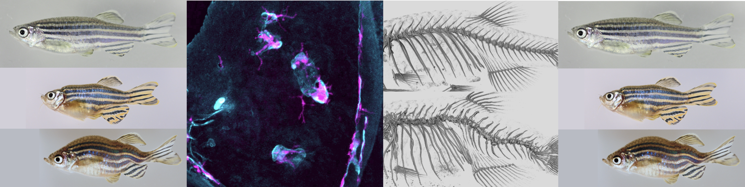
Research Focus
Our lab explores gene function during development, homeostasis and disease of the musculoskeletal system utilizing the zebrafish as a model. Zebrafish are widely used in biomedical research due to ease of genetic manipulation, high fecundity, external fertilization, rapid early development and transparency of early life stages. They show strong similarities with human physiology and in genetic architecture. While at first glance the skeleton of the zebrafish looks very different compared to a human skeleton, it contains the same major cell types and shows a strong correlation in its structure and genetic regulation. Taking advantage of the many imaging, genetic and genomic tools in zebrafish and the diverse expertise in the Orthopaedics Department, we seek to gain crucial insight into musculoskeletal biology, disease etiology and progression to aid in the development of new treatments and therapeutic strategies.

WT, heterozygous and homozygous dmh18 mutant zebrafish with deformations in the dermal and craniofacial skeleton.
Genetics of craniofacial dismorphologies
This project takes advantage of mutants with craniofacial deformations identified in a forward genetic screen to identify the genes that play a role in craniofacial development and disease. One of these mutants shows skeletal defects similar to phenotypes seen in patients with craniosynostosis. Early fusion of the cranial sutures, craniosynostosis, is the second most common craniofacial birth defect (1/2500 births). We are phenotypically, mechanistically and genetically characterizing this mutant, with the goal to elucidate the pathomechanisms of craniosynostosis to develop novel, non-surgical treatment strategies.

Remodeling of the operculum by osteoclasts. Osteoclasts in magenta, bone in cyan.
Skeletal Homeostasis - Clastokines
The skeleton is constantly remodeled through the action of bone forming osteoblasts and bone resorbing osteoclasts. To maintain a healthy, functional skeleton, both processes need to be in perfect balance which requires communication between osteoblasts and osteoclasts. While many factors are known that signal from osteoblasts to osteoclasts, less is known about Clastokines, the factors that are used by osteoclasts to communicate with osteoblasts. Utilizing transgenic zebrafish that express fluorescent markers in different populations of skeletal cells, we can image cell behavior and isolate individual cells for analysis. Through comparison of gene expression levels between functional and defective osteoclasts, we aim to identify Clastokines that control osteoblast activity and function.

Wild-type zebrafish (top) and mutant models of osteogenesis imperfecta (middle) and idiopathic scoliosis (bottom).
Forward Genetics
Forward genetic screens are an important tool for the unbiased identification of genes that play a role in developmental processes. Large-scale screens in zebrafish have been instrumental in defining the molecular basis of many aspects of development. We have isolated a large number of mutants in a screen for phenotypic changes in the form of the skeleton. Genetic mapping of the underlying gene mutations revealed that many of the identified mutants harbored changes in genes associated with human disease. Detailed phenotypic analysis of these mutants gains new insight into disease pathology and identifies new disease mechanisms. In addition these disease models are used to find new treatment strategies.